Posts

Showing posts from April, 2025

Retinal Vein Occlusion

Image
  🩸 Retinal Vein Occlusion (RVO) A common vascular disorder of the retina resulting in vision loss due to blockage of retinal venous outflow. 🔍 Definition Retinal vein occlusion occurs when a retinal vein becomes blocked , leading to venous stasis, hemorrhage , and edema of the retina , especially the macula . 🧠 Types of RVO Type Description Central Retinal Vein Occlusion (CRVO) Obstruction of the central retinal vein at the optic nerve Branch Retinal Vein Occlusion (BRVO) Obstruction of a smaller branch vein , usually at an arteriovenous (AV) crossing Hemispheric RVO Affects either the superior or inferior half of the retina 📊 Risk Factors Hypertension (most common) Diabetes mellitus Hyperlipidemia Glaucoma Smoking Hypercoagulable states (e.g., Protein C/S deficiency, antiphospholipid syndrome) Older age (>50) Oral contraceptives , especially in young women ⚙️ Pathophysiology Vein compression (at AV crossing) → Turbulent flow →...

Vitreous Hemorrhage

Image
  🩸 Vitreous Hemorrhage Bleeding into the vitreous cavity — the clear, gel-like substance that fills the eye behind the lens and in front of the retina. 🔍 Definition Vitreous hemorrhage is the presence of blood within the vitreous humor , caused by bleeding from retinal vessels, neovascularization, trauma, or other retinal pathologies. 🔬 Anatomy Recap The vitreous body is avascular (no blood vessels). Hemorrhage usually results from blood entering from adjacent structures , especially the retina , optic disc , or ciliary body . 🧠 Causes 👁️‍🗨️ Common Causes: Category Examples Retinal Neovascularization ➤ Diabetic Retinopathy (most common) ➤ Retinal vein occlusion ➤ Sickle cell retinopathy Trauma ➤ Blunt or penetrating trauma ➤ Post-surgical (cataract, vitrectomy) Posterior Vitreous Detachment (PVD) ➤ May tear a retinal vessel Retinal Tears/Detachment ➤ Associated with bleeding Others ➤ Terson's syndrome (subarachnoid hemorrhage) ➤ Blood dyscrasias (e.g., l...

Open-Angle Glaucoma

Image
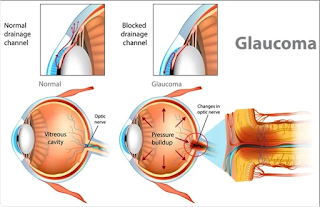
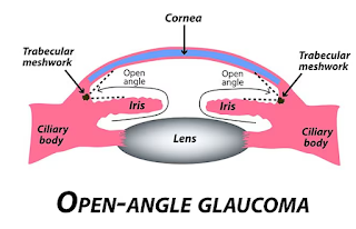
👁️‍🗨️ Open-Angle Glaucoma (OAG) Also known as Primary Open-Angle Glaucoma (POAG) — the most common type of glaucoma worldwide. 🧠 Definition Open-angle glaucoma is a chronic, progressive optic neuropathy caused by increased resistance to aqueous outflow through the trabecular meshwork , leading to elevated intraocular pressure (IOP) and gradual damage to the optic nerve — even though the anterior chamber angle remains anatomically open . 🔬 Pathophysiology Aqueous humor flows from the ciliary body → posterior chamber → pupil → anterior chamber → drains via trabecular meshwork → Schlemm’s canal → episcleral veins . In OAG: The drainage system becomes inefficient over time. Intraocular pressure rises , damaging the retinal ganglion cells and optic nerve fibers , particularly in the superior and inferior poles , leading to cupping and visual field loss . Despite the name, the angle remains open on gonioscopy. 📊 Epidemiology Most common in individ...

Angle-Closure Glaucoma

Image
👁️ Angle-Closure Glaucoma (ACG) Also known as Closed-Angle Glaucoma or Narrow-Angle Glaucoma 🔍 Definition Angle-closure glaucoma is a type of glaucoma where the anterior chamber angle between the cornea and iris is narrow or closed , impeding the outflow of aqueous humor , resulting in a sudden or chronic rise in intraocular pressure (IOP) and optic nerve damage . 🔬 Classification 1. Acute Angle-Closure Glaucoma (AACG) Ophthalmic emergency Sudden onset with severe symptoms Rapid IOP rise 2. Chronic Angle-Closure Glaucoma Gradual closure of the angle Often asymptomatic until vision is affected 3. Intermittent Angle Closure Transient symptoms Triggered by low light, emotional stress, or drugs that dilate the pupil ⚙️ Pathophysiology Aqueous humor is produced by the ciliary body → flows from posterior chamber → through pupil → anterior chamber → drains via trabecular meshwork in the angle . In angle-closure, the iris bows forward and ...

Congenital Glaucoma

Image
👶 Congenital Glaucoma (Pediatric Glaucoma) Also known as Primary Congenital Glaucoma (PCG) or Infantile Glaucoma 🧠 Definition Congenital glaucoma is a rare condition present at birth or within the first few years of life, caused by abnormal development of the eye’s aqueous outflow system , especially the trabecular meshwork and anterior chamber angle , leading to increased intraocular pressure (IOP) . 🔍 Types of Congenital Glaucoma Primary Congenital Glaucoma Isolated developmental abnormality Bilateral in 70–80% of cases Most common form of glaucoma in infancy Secondary Congenital Glaucoma Associated with ocular or systemic conditions like: Axenfeld-Rieger syndrome Peter’s anomaly Sturge-Weber syndrome Aniridia Infantile and Juvenile Glaucoma Infantile: onset after 1 month but before 3 years Juvenile: onset after age 3 to early adulthood 🧬 Etiology & Pathogenesis Developmental anomaly of the trabecular meshwork a...